|  | |
 A Mutation occurs when a DNA gene is damaged or changed in such a way as to alter the genetic message carried by that gene.A Mutagen is an agent of substance that can bring about a permanent alteration to the physical composition of a DNA gene such that the genetic message is changed. A Mutation occurs when a DNA gene is damaged or changed in such a way as to alter the genetic message carried by that gene.A Mutagen is an agent of substance that can bring about a permanent alteration to the physical composition of a DNA gene such that the genetic message is changed.Once the gene has been damaged or changed the mRNA transcribed from that gene will now carry an altered message. The polypeptide made by translating the altered mRNA will now contain a different sequence of amino acids. The function of the protein made by folding this polypeptide will probably be changed or lost. In this example, the enzyme that is catalyzing the production of flower color pigment has been altered in such a way it no longer catalyzes the production of the red pigment. No product (red pigment) is produced by the altered protein.In subtle or very obvious ways, the phenotype of the organism carrying the mutation will be changed. In this case the flower, without the pigment is no longer red. | ||
| Mutagens | ||
Chemical Mutagens change the sequence of bases in a DNA gene in a number of ways;
Radiation can also cause double strand breaks in the DNA molecule, which the cell's repair mechanisms cannot put right. Sunlight contains ultraviolet radiation (the component that causes a suntan) which, when absorbed by the DNA causes a cross link to form between certain adjacent bases. In most normal cases the cells can repair this damage, but unrepaired dimers of this sort cause the replicating system to skip over the mistake leaving a gap, which is supposed to be filled in later. Unprotected exposure to UV radiation by the human skin can cause serious damage and may lead to skin cancer and extensive skin tumors. Spontaneous mutations occur without exposure to any obvious mutagenic agent. Sometimes DNA nucleotides shift without warning to a different chemical form (know as an isomer) which in turn will form a different series of hydrogen bonds with it's partner. This leads to mistakes at the time of DNA replication. | ||
THIS IS BIOLOGY MY DEAR

Wednesday, 11 September 2013
WHAT IS MUTATION?
CELL AND FUNCTION
Cell Structure and Function
Organelles and Their Functions
In this lab you will look at the eukaryotic cells of plants and animals. Eukaryotic cells are distinguished from the more primitive prokaryotic cells by the presence of 1) cytoplasmic membranous organelles, 2) a nuclear membrane (i.e. a true nucleus), and 3) chromosomal proteins. In this lab we will focus primarily on organelles, their functions within the cell and how they differ between plant and animal cells.
Think of the cell as a microscopic city. Like a real city it requires many services to keep it clean and running smoothly. Think of some of the services a real city needs: traffic control, waste disposal, and authority figure just to name a few. Like our imagined city a cell needs the same services. Organelles are the “workers” that provide these services. The following is a list describing the various functions of some common organelles.
The NUCLEUS (“mayor of city hall”)
The nucleus houses the majority of genetic material of a cell. The nucleus is the “brain” of the cell and controls all activity within the cell. Using DNA as a blueprint
(like the blueprints of a city) the nucleus directs the production of proteins. You will learn about this process in the DNA Transcription and Translation lab.

A nucleus with the DNA coiled into chromatin. Electron microscope picture of a nucleus

RIBOSOMES (“lumber or brick yard”)
The ribosomes carry out manual labor in the form of protein synthesis for the nucleus. They bring together all the raw ingredients such as RNA (copies of the original DNA blueprints) and amino acids to assemble proteins. The proteins created are essential to cell and organismal function. Think of proteins as machinery for cell functions much like electricity and plumbing are essential in a real city. For example, enzymes are a type of protein without which life could not exist.
The large and small subunits of ribosomal RNA translating an mRNA strand into a polypeptide chain.
Refer to DNA Transcription and Translation for further reading.

The ENDOPLASMIC RETICULUM (“highways and road systems”)
There are two types of endoplasmic reticulum (ER) – Smooth ER and Rough ER. This extensive network makes up approximately one half of all membranous tissue of the cell and is the site of membrane and protein synthesis. The ER system is much like a road system along which industry can be found. Goods are manufactured and shipped to needed areas via the road system. Rough ER is named for the presence of ribosomes along its membrane and is the source of proteins. Smooth ER lacks ribosomes and is responsible for lipid synthesis and processes a variety of metabolic processes such as drug detoxification.
Can you tell the difference between the smooth and rough ER?

CELL MEMBRANE (“City Border”) and CELL WALL (“City Wall”)
Cell membranes are found in animal cells whereas cell walls are found in plant cells. Cell walls and membranes have similar functions. Like a city perimeter, cell membranes surround the cell and have the ability to regulate entrance and exit of substances, thereby maintaining internal balance. These membranes also protect the inner cell from outside forces. Cell walls, as the city analogy implies, are much stronger than cell membranes and protect cells from lysing (exploding) in extremely hypotonic (diluted) solutions. You will learn more about these concepts in the Biological Membranes lab.
Artist rendition of an animal cell membrane. Artist rendition of a plant cell wall.

CYTOSKELETON (“steel girders”)
The cytoskeleton makes up the internal framework, like the steel girders that are the framework for buildings in a city that gives each cell its distinctive shape and high level of organization. It is important for cell movement and cell division (mitosis).
Picture of a cell’s cytoskeleton- a complex network of tubules and filaments.

CYTOPLASM (“lawns and parks”)
Cytoplasm is a semi-fluid substance (think gelatin) found inside the cell. The cytoplasm encases, cushions and protects the internal organelles. It is the cell landscape found in any space where organelles are not and therefore is much like the lawns and parks of our city.
The cytoplasm is the substance surrounding the visible vacuoles in this cell.

GOLGI APPARATUS (“post office”)
Like a post office, the golgi apparatus is used for shipping those goods created by the ER and ribosomes to the rest of cell.
EM picture of a golgi apparatus Artist rendition of the Golgi Complex

CHLOROPLASTS (“solar energy plant”)
Chloroplasts are organelles found only in plant cells. Like a solar energy plant they use sunlight to create energy for the city. Chloroplasts are the site of photosynthesis a process in which the plant uses carbon dioxide, water and sunlight to create energy in the form of glucose for the plant cell as well as heterotrophs that consume the plant.
Artist rendition of a chloroplast- site of photosynthesis in plant cells.

MITOCHONDRIA (“energy plant”)
Mitochondria are found in both plant and animal cells and is the site of cellular respiration. Through this process that will be covered in the Photosynthesis and Respiration lab ATP is created which is used for energy by the cell.
Electron microscope picture of a mitochondria.

LYSOSOMES (“waste disposal and recycling”)
The lysosomes are digestive sacs that can break down macromolecules in the cell using the process of hydrolysis. The digestion is carried out with lysosomal enzymes found in the lysosome. Like waste disposal in a city, lysosomes help keep excessive or bulky macromolecules from building up in the cell.
Electron microscope picture of a lysosome.

VACUOLES and VESICLES (“warehouses, water towers or garbage dumps”)
Think of these membrane sacs that have a variety of functions as containment units for anything in excess in a city. They can hold many substances from organic molecules to simple excess water. Plant cells have a central vacuole that is important in maintaining plantturgidity. You can read more about this phenomenon in the Biological Membranes Lab.
Central vacuole of a plant cell.

Plant versus Animal Cells
Now that you know some important cell organelles let us identify those that distinguish plant cells from animal cells. From the descriptions above, we can identify three organelles unique to plant cells: 1) cell wall (versus a cell membrane in animal cells), 2) central vacuole (regular vacuoles are found in animal cells) and 3) chloroplasts (animals do not perform photosynthesis. This is what makes plants autotrophs and animals heterotrophs.)
| CELL STRUCTURE | LOCATION | DESCRIPTION | FUNCTION |
Cell Wall
 |
Plant, Fungi, & Bacteria, but not animal cells
|
|
|
Cell Membrane |
All cells
|
|
|
Nucleus
 |
All cells except prokaryotes
|
|
|
Nuclear membrane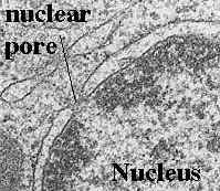 | All cells except prokaryotes |
|
|
Cytoplasm
 |
All cells
|
|
|
| Endoplasmic reticulum (ER)  | All cells except prokaryotes |
|
|
Ribosome |
All cells
|
|
|
Mitochondrion |
All cells except prokaryotes
|
|
|
Vacuole |
Plant cells have a single, large vacuole
Animal cells have small vacuoles |
|
|
Lysosome |
Plant - uncommon
Animal - common |
|
|
Chloroplast |
Plants and algae
|
|
|
nucleolus |
All cells except prokaryotes
|
|
|
Golgi Apparatus
|
All cells except prokaryotes
|
|
|
Cilia |
Animal cells, Protozoans
|
|
|
Flagellum |
Bacterial cells & Protozoans
|
|
|
Centrioles |
Animal cells
|
|
|
Cytoskeleton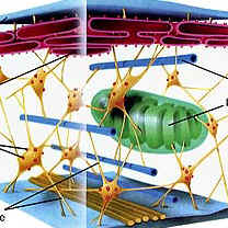 |
All cells
|
|
|
MITOSIS AND MEIOSIS
Mitosis and Meiosis
What is Mitosis?
Mitosis produces two daughter cells that are identical to the parent cell. If the parent cell is haploid (N), then the daughter cells will be haploid. If the parent cell is diploid, the daughter cells will also be diploid.
N → NThis type of cell division allows multicellular organisms to grow and repair damaged tissue.2N → 2N
Click here to go to the chapter on Mitosis.
Summary of the Phases of Mitosis
The drawings below show chromosome movement and alignment in a cell from a species of animal that has a diploid number of 8. As you view the drawings, keep in mind that humans have a diploid number of 46.
Interphase (G1 and G2)
Chromosomes are not easily visible because they are uncoiled.Prophase The chromosomes begin to coil.
The spindle apparatus begins to form as centrosomes move apart.Prometaphase
The nuclear membrane disintegrates.
Kinetochores form on the chromosomes.
Kinetochore microtubules attach to the chromosomes.Metaphase
The chromosomes become aligned on a plane.Anaphase
The chromatids separate (The number of chromosomes doubles).Telophase
The nuclear membrane reappears.
The chromosomes uncoil.
The spindle apparatus breaks down.
The cell divides into two.G1 Interphase
The chromosomes have one chromatid.G2 Interphase
The chromosomes are replicated. Each one has two sister chromatids.


What is Meiosis?
Meiosis produces daughter cells that have one half the number of chromosomes as the parent cell.
Meiosis enables organisms to reproduce sexually. Gametes (sperm and eggs) are haploid.2N → N
Meiosis involves two divisions producing a total of four daughter cells.
Summary of the Phases of Meiosis
A cell undergoing meiosis will divide two times; the first division is meiosis 1 and the second is meiosis 2. The phases have the same names as those of mitosis. A number indicates the division number (1st or 2nd):
meiosis 1: prophase 1, metaphase 1, anaphase 1, and telophase 1In the first meiotic division, the number of cells is doubled but the number of chromosomes is not. This results in 1/2 as many chromosomes per cell.
meiosis 2: prophase 2, metaphase 2, anaphase 2, and telophase 2
The second meiotic division is like mitosis; the number of chromosomes does not get reduced.
Prophase IHomologous chromosomes become paired.
Crossing-over occurs between homologous chromosomes.
Crossing over Metaphase IHomologous pairs become aligned in the center of the cell.
The random alignment pattern is called independent assortment. For example, a cell with 2N = 6 chromosomes could have any of the alignment patterns shown at the left.. Anaphase IHomologous chromosomes separate. Telophase IThis stage is absent in some species InterkinesisInterkinesis is similar to interphase except DNA synthesis does not occur. Prophase II Metaphase II Anaphase II Telophase II Daughter Cells


Laboratory Exercise
Mitosis in Animals
Human Chromosomes
1. View a slide of human chromosomes and draw some of the chromosomes below.2. Do the chromosomes have one chromatid or two?Below: Human Chromosomes Click on the photograph to view an enlargement.Whitefish Blastula
3. The cells of a developing embryo are dividing rapidly and can be used for viewing the different stages of mitosis. Obtain a whitefish blastula (early embryo) slide and find a cell in each of these phases: interphase, prophase, metaphase, anaphase, and telophase.4. Draw a cell in anaphase below.Below: Whitefish blastula mitosis X 400. Click on the images to view enlargements.Additional Photographs of mitosis in animals: Ascaris Megalocephala Mitosis


Mitosis in Plants
1. Cells at the tips of plant roots and stems grow rapidly and can be used for viewing the stages of mitosis. Used a slide of onion (Allium) root tip to identify interphase, prophase, metaphase, anaphase, and telophase.2. Draw a cell in telophase below. Draw the cell plate if it is visible.Below: Allium root tip mitosis X 400. Click on the images to view enlargements.


Meiosis in Animals (Gametogenesis)
Testis
1. Obtain a slide of a cross section of rat testes and observe the seminiferous tubules. Identify spermatogonia. Identify sperm.Below: Rat testes X 100. Click on the image to view an enlargement.Ovary
The primary oocyte is contained within a structure called a follicle. As the follicle enlarges, it produces hormones. During ovulation, the follicle ruptures and releases the secondary oocyte.2. View a slide of a section of a rabbit ovary under scanning magnification and observe follicles in various stages of development. Can you see an oocyte in any of the follicles?Below: Rabbit ovary X 40. Click on the image to view an enlargement.


Subscribe to:
Posts (Atom)